FIGURE 2.

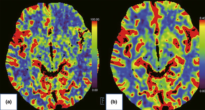
Computed tomography brain perfusion imaging. (a) Preoperative impaired left anterior frontal lobe, the left temporal lobe, and the left basal ganglia blood flow (blue). (b) Postoperative improvement

Computed tomography brain perfusion imaging. (a) Preoperative impaired left anterior frontal lobe, the left temporal lobe, and the left basal ganglia blood flow (blue). (b) Postoperative improvement