
FIGURE 2.
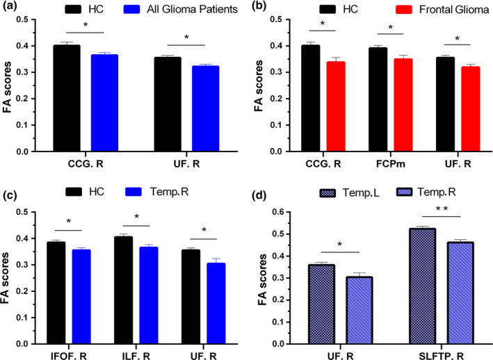
Comparisons of fractional anisotropy (FA) between HCs and all patients with glioma (a), HCs and patients with glioma located in the frontal lobe (b), HCs and patients with glioma located in the right temporal lobe (c), and patients with glioma located in the left temporal lobe and patients with glioma located in the right temporal lobe (d). Notes: Data are expressed as the mean FA value (error bars representing standard error of the mean). *p < .05, **p < .01. No significance was found between HCs and patients with gliomas located in the left temporal lobe. CCG.R, right cingulum (cingulate gyrus); FCPm, forceps minor; IFOF.R, right inferior fronto‐occipital fasciculus; ILF.R, right inferior longitudinal fasciculus; SLFTP.R, right superior longitudinal fasciculus temporal part; Temp.L/R, patients with glioma located in the left/right temporal lobe; UF.R, right uncinate fasciculus