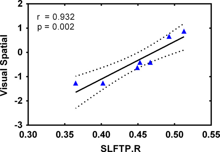
FIGURE 3.

Association of significantly altered FA values of SLFTP.R and declined visual–spatial measures. Notes: ▲ represents values of patients with glioma located in the right temporal lobe. Four patients did not complete all cognitive tests. The solid line and dashed lines represent the best‐fit line and 95% confidence interval of partial correlation, respectively. SLFTP.R, right superior longitudinal fasciculus temporal part.
