FIGURE 1.

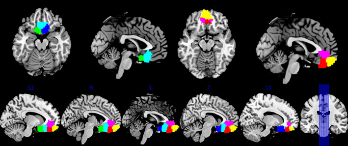
Locations of the six‐cluster solution within the vmFL (clusters color‐coded as follows: 1 = blue, 4 = cyan, 6 = green; 2 = yellow, 3 = magenta, 5 = red). vmFL, ventromedial regions of the frontal lobe

Locations of the six‐cluster solution within the vmFL (clusters color‐coded as follows: 1 = blue, 4 = cyan, 6 = green; 2 = yellow, 3 = magenta, 5 = red). vmFL, ventromedial regions of the frontal lobe