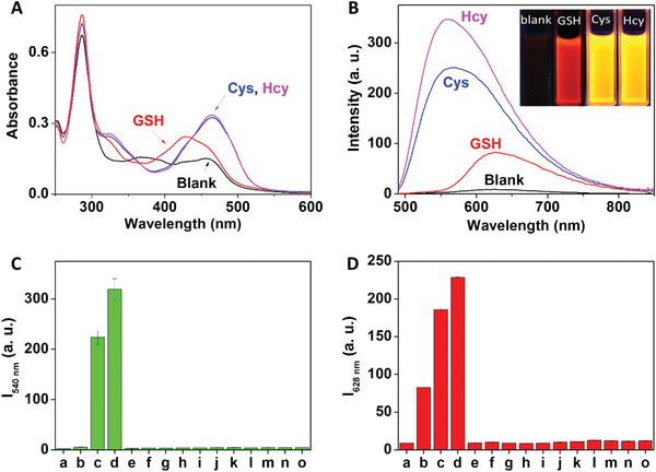
Figure 1.

Spectrometric response of Ru‐NBD to biothiols. A) Absorption and B) steady‐state emission changes of Ru‐NBD (10 × 10−6 m) in the absence and the presence of GSH, Cys, and Hcy in 50 × 10−3 m Tris‐HCl buffer of pH 7.4 (B inset: photo of luminescence color changes of Ru‐NBD in the absence and presence of GSH, Cys and Hcy). Emission intensity of Ru‐NBD (10 × 10−6 m) at C) 540 nm and D) 628 nm after reacted with different amino acids (200 × 10−6 m) in 50 × 10−3 m Tris‐HCl buffer of pH 7.4. Amino acids: a) blank, b) GSH, c) Cys, d) Hcy, e) tryptophan, f) threonine, g) glycine, h) valine, i) leucine, j) histidine, k) proline, l) serine, m) tyrosine, n) alanine, and o) aspartic acid.
