
Figure 1.
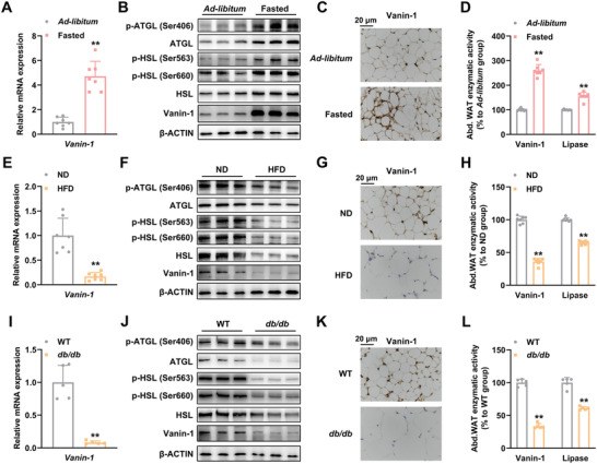
Vanin‐1 functions as a nutrient‐sensitive factor in mouse abdominal WAT. For (A)–(D), mice were subjected to either 24 h fasting or ad libitum. A) RT‐qPCR analysis of Vanin‐1 mRNA level in the abdominal WAT. B) Western blot analysis of Vanin‐1 and key lipolytic protein expression in the abdominal WAT. C) IHC analysis of Vanin‐1 expression in the abdominal WAT. D) Enzymatic activities of Vanin‐1 and total lipase in the homogenates of abdominal WAT. ** P < 0.01 versus ad libitum group. n = 7. For (E)–(H), mice were fed with either a normal diet (ND) or an HFD for 16 weeks. E) RT‐qPCR analysis of Vanin‐1 expression in the abdominal WAT. F) Western blot analysis of Vanin‐1 and key lipolytic protein expression in the abdominal WAT. G) IHC analysis of Vanin‐1 expression in the abdominal WAT. H) Enzymatic activities of Vanin‐1 and total lipase in the homogenates of abdominal WAT. ** P < 0.01 versus ND group. n = 7. I) RT‐qPCR analysis of Vanin‐1 mRNA levels in the abdominal WAT from WT or db/db mice. J) Western blot analysis of Vanin‐1 and key lipolytic protein expression in the abdominal WAT from WT or db/db mice. K) IHC analysis of Vanin‐1 expression in the abdominal WAT from WT or db/db mice. L) Enzymatic activities of Vanin‐1 and total lipase in the homogenates of abdominal WAT from WT or db/db mice. ** P < 0.01 versus WT group. n = 5. All values are presented as the mean ± SD. Unpaired Student's t‐test was used for comparison between two groups. Abd. WAT: abdominal WAT.