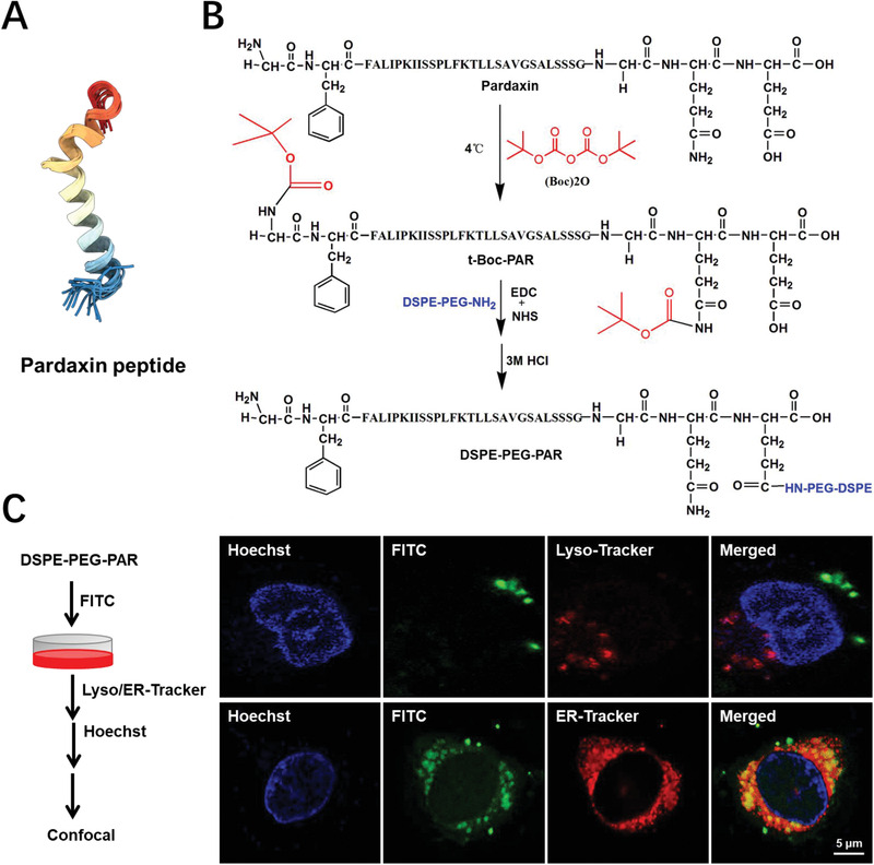
Figure 1.

The synthesis and intracellular localization of DSPE‐PEG‐PAR after 2 h incubation with MCF‐7 cells. A) The spatial structure of the pardaxin peptide. The structure image was obtained from the RCSB Protein Data Bank (https://www.rcsb.org/). B) The organic synthetic routes of DSPE‐PEG‐PAR. C) The intracellular co‐localization images of DSPE‐PEG‐PAR with lysosomes and ER were obtained by confocal microscopy, bar: 5 µm.
