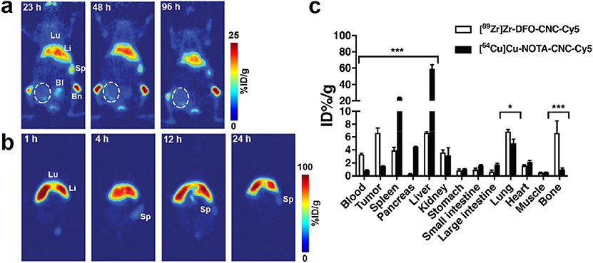
Fig. 3.
a) Serial PET imaging of the same animal bearing single orthotopic 4T1 mammary fat pad allograft after intravenous administration of 3.7 MBq (60 μg) of [89Zr]Zr-DFO-CNC-Cy5 shows accumulation of the nanocrystals in the liver, spleen, and bone. The tumor area (circled) shows only slight radioactive signal on the perimeter of the tumor. b) Serial PET imaging of a single mouse after intravenous administration of 7.4 MBq (60 μg) of [64Cu]Cu-NOTA-CNC-Cy5 shows rapid accumulation of the tracer in the liver, preventing delineation of the tumor in the image. Lu, lung; Li, liver; Sp, spleen; Bl, bladder; Bm, bone with marrow. PET images were acquired under isoflurane anesthesia on a Focus 120 Micro-PET small animal PET scanner with the imaging protocol set to register a minimum of 20 million coincidence events. c) Ex vivo biodistribution of [89Zr]Zr-DFO-CNC-Cy5 and [64Cu]Cu-NOTA-CNC-Cy5 at 24 h p.i. shows significant differences in the biodistribution of the two materials. Columns denote mean ± SD of n = 3. Statistical analysis was carried out with an unpaired t-test with the two-stage step-up method of Benjamin, Krieger and Yekutieli with the false discovery rate set to Q = 10%; *p < 0.05, **p < 0.01, and ***p < 0.001.

