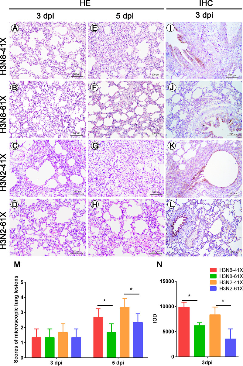
FIG 6.
Histopathological changes and IHC staining in the lungs of dogs infected with CIVs. Hematoxylin and eosin staining of lung sections from dogs infected with H3N8-41X (A and E), H3N8-61X (B and F), H3N2-41X (C and G), or H3N2-61X (D and H) at 3 dpi (A, B, C, and D) or 5 dpi (E, F, G, and H). Microscopic lesions were counted (M). IHC staining of lungs of dogs infected with H3N8-41X (I), H3N8-61X (J), H3N2-41X (K), or H3N2-61X (L) was performed using an anti-NP antibody, and the results are presented as IOD values from five random fields (N). Values represent means and standard deviations. *, P < 0.05.

