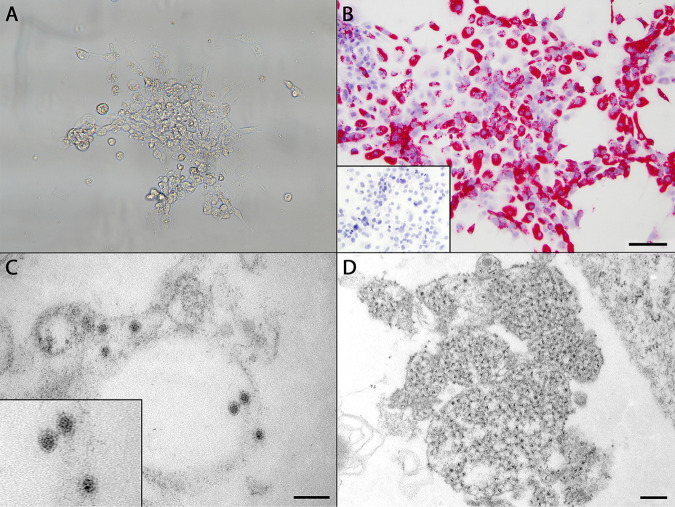
FIG 2.
(A) Cytopathic effect in SFV-infected striped-snakehead (SSN-1) cells incubated at 20°C, beginning with mild vacuolation and rounding. By day 10 postinoculation, all cells had lifted from the flask or lysed. (B) RNAscope in situ hybridization assay demonstrating strong positive cytoplasmic staining of SFV-infected SSN-1 cells by Vulcan Fast Red. Bar, 100 μm. (Inset) Lack of staining in uninoculated control cells. (C) Transmission electron microscopic image of SFV prepared from infected SSN-1 cells. Viral particles were roughly round, with an average diameter of 35.17 nm, and possessed an electron-dense core surrounded by an outer membrane. Particles were present primarily within the cisternae of the endoplasmic reticulum and in vesicles. Bar, 100 nm. (Inset) Enlarged image of three viral particles with typical flaviviral morphology. (D) Cytoplasmic replication complex of SFV within infected SSN-1 cells filled by ill-defined, uncondensed material believed to represent early RNA synthesis. Bar, 200 nm.

