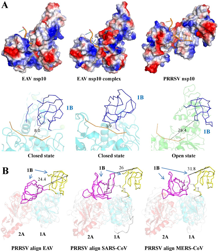
FIG 5.
Dynamic conformational states of the 1B domain in arterivirus helicases. (A) The structures of PRRSV nsp10 and EAV nsp10 (PDB ID 4N0N and 4N0O) are shown and are colored according to electrostatic potential as described in Fig. 1D. The ssDNA from the EAV nsp10 complex (PDB ID 4N0O) was docked into PRRSV nsp10 and EAV nsp10 (PDB ID 4N0N). The distance between the ssDNA substrate and the 1B domain was measured by using PyMOL software. (B) Structural alignment between PRRSV and other nidovirus helicases (EAV nsp10, SARS-CoV nsp13, and MERS-CoV nsp13). The PDB ID is consistent with Fig. 3A.

