Table 1:
Summary of Optoelectronic Devices
| Device: | Type: | How it works: | Benefits: | Limitations: |
|---|---|---|---|---|
ARGUS II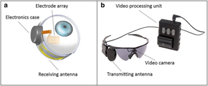 Source: Courtesy of SecondSight |
Epiretinal implant with external camera | External camera records and transmits pulses wirelessly through microelectrode array to inner retina | Unaffected by corneal or lens opacities due to wireless transmission from camera to array; long-term safety has been demonstrated | Visual perception through fixed camera is independent of eye movement; 60 electrodes limits potential visual acuity |
IRIS II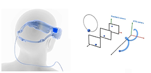 Source: Courtesy of Pixium Vision |
Epiretinal implant with external camera | External camera records and transmits pulses wirelessly through microelectrode array to inner retina | Exchangeable system allows for replacement; unaffected by corneal or lens opacities due to wireless transmission from camera to array | Visual perception through fixed camera is independent of eye movement; 150 electrodes but requires further studies to demonstrate efficacy |
Alpha IMS/AMS (Discontinued)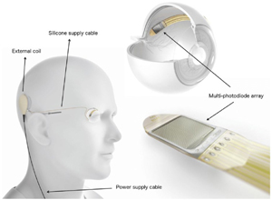 Source: Retina Implant AG, Reutlingen, Germany, used by Bloch et al. 2019, licensed under CC-NY. |
Subretinal implant | Photodiode array in subretinal space that detects light and converts it to electrical signals transmitted to overlying retina | Vision generated corresponds with natural eye movement due to subretinal implant itself directly detecting light | Affected by opacities between cornea and retina due to requiring light to reach the retina; 1500 (IMS) or 1600 (AMS) electrodes but visual acuity significantly below potential |
PRIMA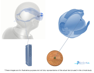 Source: Courtesy of Pixium Vision |
Subretinal implant with external camera | External camera records video that is processed and projected via infrared laser patterns through the pupil to subretinal implant, where photovoltaic array converts the light to electrical signals stimulating bipolar cells | Design of subretinal implant includes ground grid around each stimulating electrode allowing theoretical improvement of resolution | Affected by opacities between cornea and retina due to requiring light to reach the retina; 378 pixels (electrodes) but requires clinical trials to demonstrate efficacy |
Bionic Vision Australia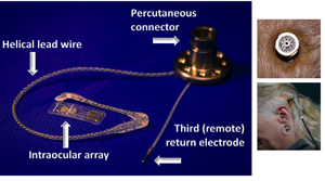 Source: Courtesy of Ayton et al. 2014, originally from Bionic Vision Technologies |
Suprachoroidal implant with external camera | External camera records and transmits pulses wirelessly through microelectrode array through choroid to inner retina | Unaffected by corneal or lens opacities due to wireless transmission from camera to array | Visual perception through fixed camera is independent of eye movement; 44 channels (electrodes) limits potential visual acuity and requires clinical trials to demonstrate efficacy |
Orion Cortical Implant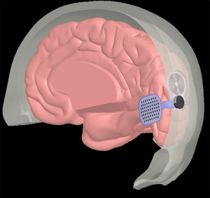 Source: Courtesy of SecondSight |
Cortical implant with external camera | External camera records and transmits pulses wirelessly to microelectrode array to medial occipital lobe | Unaffected by corneal or lens opacities due to wireless transmission from camera to array; potential for use in patients with significant inner retina and/or optic nerve degeneration | Visual perception through fixed camera is independent of eye movement; 60 electrodes limits potential visual acuity; requires clinical trials to demonstrate efficacy and long-term safety |
