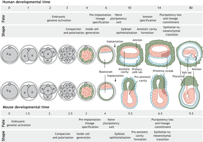
Fig. 1.
Overview of human and mouse embryo development. Upon fertilisation, mouse and human embryos undergo a series of cleavage divisions. The embryonic genome becomes activated by the two-cell stage in mouse embryos and at the four/eight-cell stage transition in human embryos. It is followed by compaction and polarisation, which occur at the eight-cell stage in mouse embryos, and between the eight- to 16-cell stage in human embryos. Formation of a hollow cavity, the blastocoel, denotes the formation of the blastocyst, which, by embryonic day E4 (mouse) and E6 (human), is composed of three main tissues: epiblast, hypoblast and trophectoderm. Upon implantation (E5 in mouse and E7 in human), embryos undergo a global morphological transformation. The embryonic epiblast loses its naïve pluripotent character, becomes epithelial and forms the pro-amniotic cavity (mouse), which spans both the epiblast and the trophectoderm-derived extra-embryonic ectoderm, and the amniotic cavity (human). A key difference between mouse and human embryos relates to the formation of the amnion. Whereas in mouse embryos amnion formation takes place during gastrulation, in human embryos a subset of epiblast cells differentiates to form the squamous amniotic epithelium during early post-implantation (E10). As a result, the human epiblast acquires a disc shape and the mouse epiblast a cylindrical morphology. On embryonic days 6.5 (mouse) and 14 (human), gastrulation is initiated in the posterior epiblast. Cells undergo an epithelial-to-mesenchymal transition, lose their pluripotent character and commit to one of the germ layers. Epiblast-derived tissues are shown in pink, hypoblast-derived tissues are shown in blue and trophectoderm-derived tissues are shown in green.

