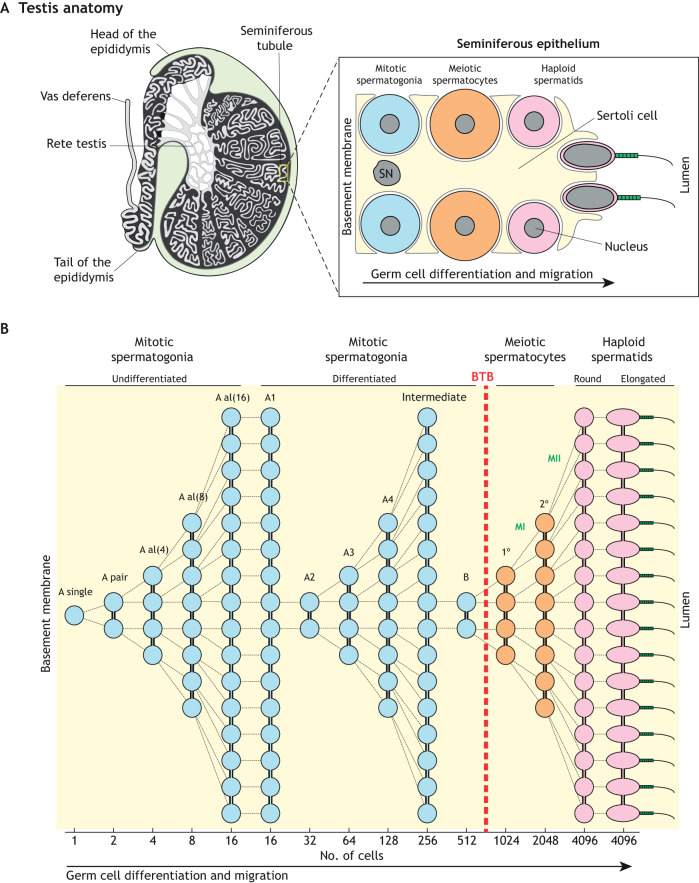
Fig. 3.
Spermatogenesis. (A) Left panel, anatomy of the mammalian testis highlighting the convoluted seminiferous tubules in which spermatogenesis takes place. Right panel, schematic illustration of the seminiferous epithelium highlighting the intimate association between somatic Sertoli cells and germ cells. For simplicity, only the major germ cell types are shown. (B) Cellular pedigree of a single undifferentiated spermatogonium, highlighting germ cell amplification. The theoretical number of syncytial cells at each stage is shown at the bottom. Note that meiotic spermatocytes and post-meiotic spermatids develop on the adluminal side of the blood-testis barrier (BTB). SN, Sertoli cell nucleus; A al, A aligned; 1°, primary spermatocyte; 2°, secondary spermatocyte; MI, meiosis I; MII, meiosis II.

