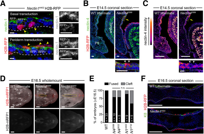
Fig. 2.
Depletion of one Nectin homolog is insufficient to cause highly penetrant CP. (A) E14.5 Nectin14402-transduced palatal epithelia immunostained for nectin 1 (green) and H2B-RFP reporter (red). Boxed regions show nectin 1 in greyscale. (B,C) Immunofluorescent images (B) and LUT intensity plots (C) of E14.5 Nectin42589 and wild-type PS showing loss of nectin 4 in H2B-RFP+ cells. Bottom panels are detailed views of the boxed regions (highlighted by yellow arrows) in the top panels. Arrowheads in the bottom panels indicate RFP+ periderm cells. (D) Dark-field (top) and fluorescent (bottom) stereoscope images of E16.5 wild-type (left), Nectin42589 (middle) and Nectin14402 (right) infected embryos, as in Fig. 1C. (E) Rate of CP penetrance in Nectin1 and Nectin4 knockdown embryos. (F) E16.5 palate in Nectin14402 embryo showing complete fusion. Scale bars: 4 µm in A; 25 µm in B,C; 1 mm in D; 100 µm in F.

