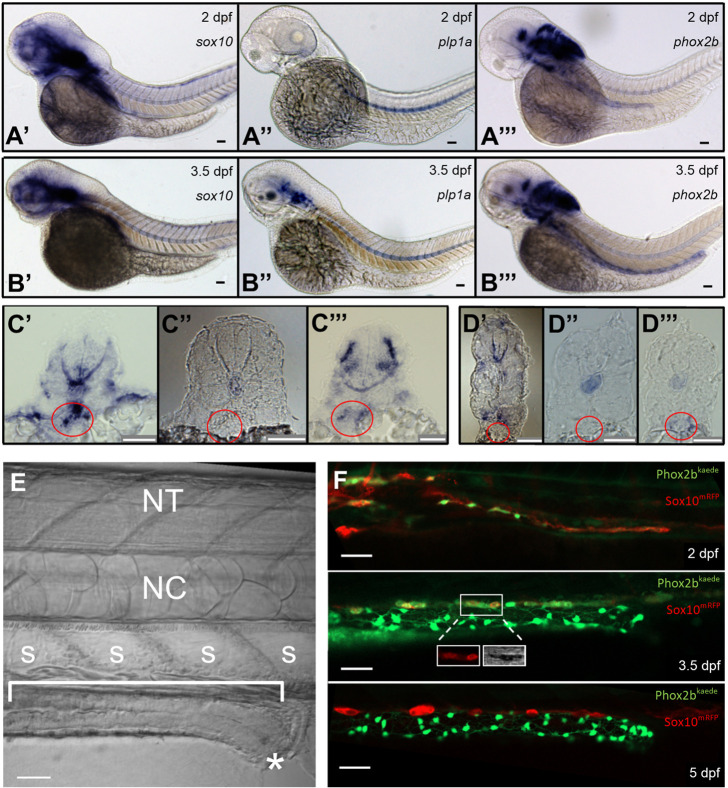
Fig. 1.
Resident neuronal progenitors are absent in the post-embryonic intestine. (A) ISH of enteric neural elements in 2 dpf embryos. sox10 is detected at 2 dpf as a stream in the midgut that does not yet extend to the hindgut (A′), plp1a exhibits no expression (A″) and phox2b has weak expression in the proximal gut (A‴). Expected probe trapping is evident in the notochord. (B) ISH of enteric neural elements in 3.5 dpf larvae. sox10 signal is absent in the intestine, although proximal probe trapping is seen in the nascent swim bladder (B′). plp1a is expressed dorsally but no expression is evident in the intestine (B″). phox2b expression extends throughout the intestine (B‴). (C′-C‴) Proximal cross-sections of 2 dpf embryos stained for sox10 (C′), plp1a (C″) and phox2b (C‴), with the nascent foregut marked by a red circle. (D′-D‴) Distal cross-sections of 3.5 dpf larvae stained for sox10 (D′), plp1a (D″) and phox2b (D‴), with the developing midgut marked by a red circle. (E) Anatomical orientation: fluorescent figures are oriented in this manner unless otherwise stated. The intestine is located ventrally (bracket) and extends anteriorly (left) to posteriorly (right), ending at the anus (*). A row of polygonal somites (S) are arranged dorsal to the intestine. The notochord (NC) and neural tube (NT, not visible in this image) are located dorsally. (F) Live imaging of Phox2b-kaede×Sox10-mRFP fish are consistent with ISH results: a migrating chain of Sox10 cells is observed in the midgut that does not yet extend to the hindgut at 2 dpf, but then Sox10 expression ceases at 3.5 dpf and 5 dpf. A few Sox10-expressing cells, seen dorsolateral to the intestine, are consistent with melanocytes, as supported by visible pigment using the transmitted light photomultiplier tube (TPMT), a detector for transmitted light (inset, 3.5 dpf panel). Scale bars: 50 µm.

