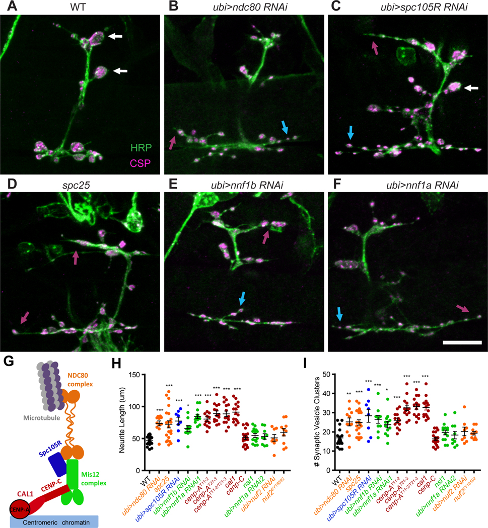
Figure 2. Many kinetochore components are required for proper NMJ formation.

(A-F) For the indicated genotypes, loss of a kinetochore protein causes overly long neurites (magenta arrows) and multiple small vesicle clusters stained with anti-CSP (blue arrows), and few or no large boutons (white arrows). (G) Schematic of the subcomplexes that make up the kinetochore. (H, I) Quantification of the phenotypes for kinetochore mutants and RNAi knockdown, colored to correspond to the subcomplexes in (G). Each genotype was compared with WT using one-way ANOVA with Dunnett’s multiple comparisons test. Scale bars, 10 μm. ***p<0.001, **p<0.01, *p<0.05. Error bars, mean ± s.e.m.
