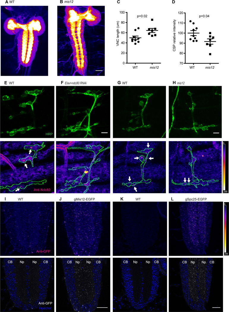
Figure 3. Kinetochore proteins localize to embryonic NMJs and synaptic neuropil.

(A, B) Anti-CSP staining of WT and mis12 mutant nervous systems. The synaptic neuropil, forming the center of the brain lobes and parallel stripes in the ventral nerve cord (VNC) is intensely stained in contrast to the surrounding cortices of cell bodies. (C, D) Quantification of VNC length (C) and anti-CSP signal intensity (D) for the genotypes in (A, B). (E-H) ndc80 antibody staining of NMJs in wildtype embryos (E, G), and in embryos expressing ndc80 RNAi in neurons (F) or in mis12f03756f03756 embryos (H). Nerve endings were identified by anti-HRP staining (top panels) and then outlined (below) in images of anti-ndc80 immunoreactivity. Sparse puncta of ndc80 (arrows) were present in boutons of wildtype embryos, but largely absent when ndc80 was knocked down by RNAi and also fewer and fainter in mis12 mutants. (I-L) Anti-GFP staining for EGFP fusion proteins of Mis12 and Spc25, in genomic constructs that place their expression under control of the endogenous promoters. In the neuropil of the embryonic ventral nerve cord Mis12-EGFP (J) and Spc25-EGFP (L) formed bright puncta of GFP immunoreactivity that were absent from embryos lacking the transgenes (I and K). CB, cell body; Np, neuropil. Scale bar, 5 μm (E-H) and 20 μm (A-D and I-L).
