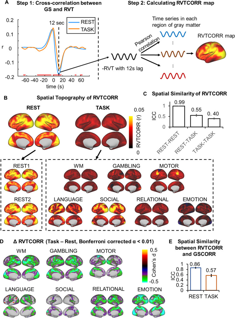
Fig 5. Spatial topography of the respiration effect.
(A) An illustration of cross-correlation between the GS and RVT, calculating a correlation map between the time series of respiration and time series in each region of the gray matter. The respiration time series was first transformed into the RVT, as the difference between the upper and lower envelope (see Materials and methods for details). Next, the cross-correlation was performed between the GS and RVT to determine the time lag with the strongest correlation, i.e., 12 second (step 1). The RVT was flipped and correlated with the time series in gray matter with 12-second lag (step 2) to obtain the RVTCORR map. The time interval showing significant rest-task differences of the GS-RVT correlations was indicated by red line, thresholded at Bonferroni-corrected α < 0.01. (B) Spatial patterns of RVTCORR in 2 days’ resting states and seven tasks. Top panel corresponded to the averaged spatial patterns for resting and task states, respectively. Bottom panel corresponds to the spatial pattern in each condition. (C) The spatial similarity (ICC ± 95% CI) of RVTCORR between the 2 days’ resting states, between the resting and task states, and between the tasks. (D) Rest-task modulation of RVTCORR. (E) Spatial similarity (ICC ± 95% CI) between GSCORR and RVTCORR for resting and task states, respectively. Data are available at Dryad: https://doi.org/10.5061/dryad.xsj3tx9bw. GS, global signal; GSCORR, GS correlation; ICC, intraclass correlation coefficient; RVT, respiration volume per time; RVTCORR, RVT correlation; WM, working memory.

