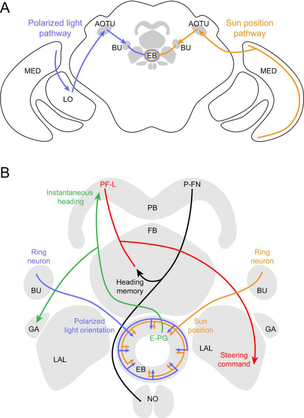
Fig. 7.

Proposed neural pathways for polarized light and sun position. (A) Polarized light (blue) and sun position (orange) are likely processed along similar pathways, illustrated here for convenience on different sides of the brain. Pathways are based on Schistocerca gregaria and, when available, D. melanogaster. Polarized light is detected by DRA photoreceptors which project to the medulla (Fortini and Rubin, 1991; Weir et al., 2016). In locusts, there are projections from the medulla to the anterior lobe of the lobula and then the lower unit of the anterior optic tubercle (AOTU; Homberg et al., 2003). From the AOTU, neurons project to the bulb (BU, the lateral triangle and median olive in locusts) and synapse onto neurons that terminate in the ellipsoid body (EB, central body lower in locusts). In flies, the polarized light pathway has not been directly characterized beyond the optic lobe but is assumed to be similar. For the sun position pathway (orange), classes of medulla neurons (TIM1, TML1) respond to a bright spot in locusts (el Jundi et al., 2011). In flies, there is a direct pathway from the medulla to the AOTU (Omoto et al., 2017), from where there are projections to the BU and then to the EB. (B) Celestial cue information is processed in the central complex and associated neuropils. Here, we indicate each columnar cell type with a single example for clarity. Ring neurons project from the BU to the EB, likely synapsing with E-PG cells (Omoto et al., 2017). E-PGs encode the fly's instantaneous heading (Seelig and Jayaraman, 2015) and respond to a sun stimulus (Giraldo et al., 2018). The E-PGs tile the EB and 16 medial glomeruli of the protocerebral bridge (PB) and project to the gall (GA), a putative output region (Seelig and Jayaraman, 2015; Wolff et al., 2015). According to an anatomically based model and electrophysiology in bees (Stone et al., 2017), flies might compare their instantaneous heading with the memory of their desired heading using P-FN cells. These P-FN neurons likely integrate input from the E-PGs and visual odometry cells (not shown) and in turn synapse onto PF-L cells (Wolff et al., 2015), which are predicted to control steering through projections to descending motor neurons (Stone et al., 2017). The outline of neuropil regions is modified from Wolff et al. (2015).
