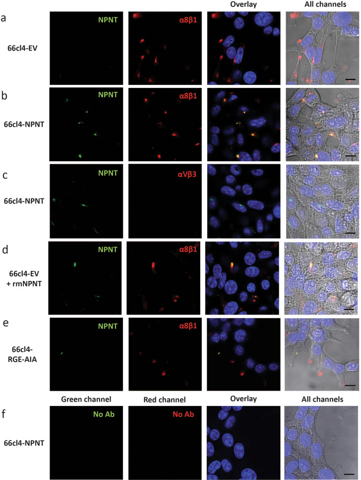
Figure 3.
NPNT co-localizes with integrin α8β1 on the 66cl4 cell surface. Immunofluorescent staining of NPNT (green), integrin subunit α8 (red) or integrin αVβ3 (red) in 66cl4 control cells or 66cl4 cells expressing either wild-type NPNT or a mutated version of NPNT (RGE-AIA). Nuclei are stained blue with dapi. (a) 66cl4-EV cells double stained for NPNT and integrin subunit α8. (b) 66cl4-NPNT cells double stained for NPNT and integrin subunit α8. (c) 66cl4-NPNT cells double stained for NPNT and integrin αVβ3.(d) 66cl4-EV cells with exogenously added rmNPNT and double stained for NPNT and integrin subunit α8. (e) 66cl4-RGE-AIA cells double stained for NPNT and integrin subunit α8. (f) Controls where 66cl4-NPNT cells were treated according to the same protocol, but with both primary antibodies omitted.

