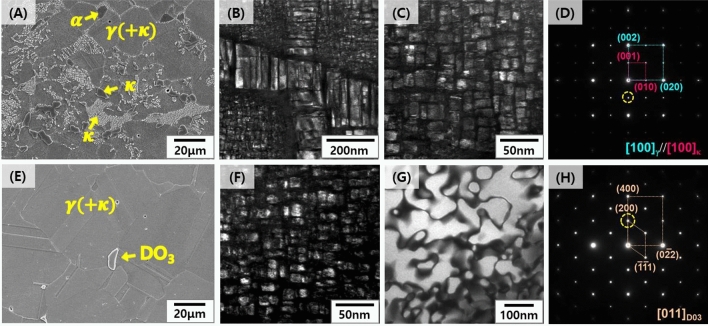
Figure 2.
Microstructures of the solution-treated samples for 2 h at 1,050°C: (A) SEM image of the LWS1 alloy (Fe–20Mn–12Al–1.5C), (B,C) dark-field TEM images of coarse intergranular κ-carbide and nano-size κ-carbide within the austenite matrix, respectively; (D) selected area diffraction (SAD) pattern of the κ-carbide; (E) SEM image of the LWSS1 alloy (Fe–20Mn–11.5Al–1.5C–5Cr); (F,G) dark-field TEM images of coarse nano-size κ-carbide within the austenite and DO3 phase, respectively; (H) SAD pattern of the DO3 phase.

