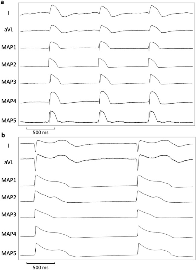
Figure 6.

Illustrative example of action potential and ECG tracings under baseline conditions (a) and after administration of erythromycin (b) in spontaneously beating bradycardic hearts under hypokalemic conditions. With erythromycin, action potentials are substantially prolonged and triangulated. Of note, spatial dispersion of repolarization (as determined by the duration differences between MAP 5 and MAP 3 in (b)) is amplified. (MAP = monophasic action potential).
