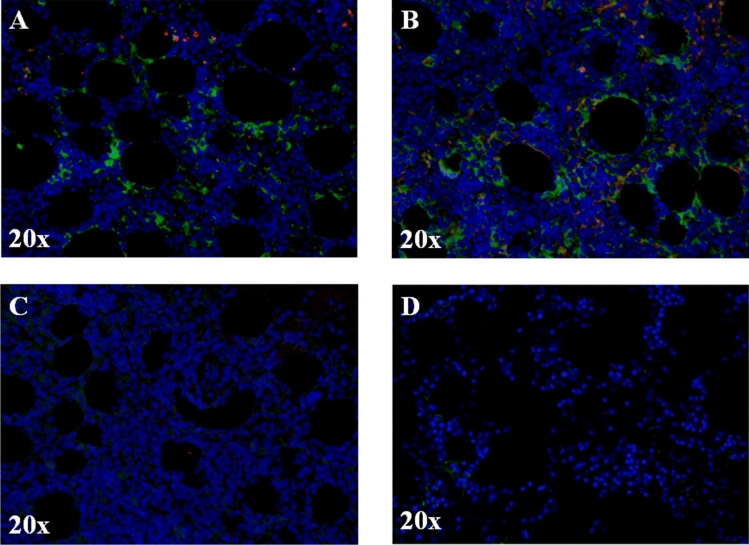
Figure 1.
FeH is more represented than FeL in BM biopsies of AOSD complicated with MAS. (A, B) Immunofluorescence staining of BM biopsies of patients with AOSD complicated with MAS. The representative images of two patients (A, B) and two HCs (C) and (D) coloured with FeH (green) and FeL (red) are shown. The intensity of FeH expression is higher, when compared with FeL, since it is more represented. Pictures are representative of all experiments. Original magnification 20×.

