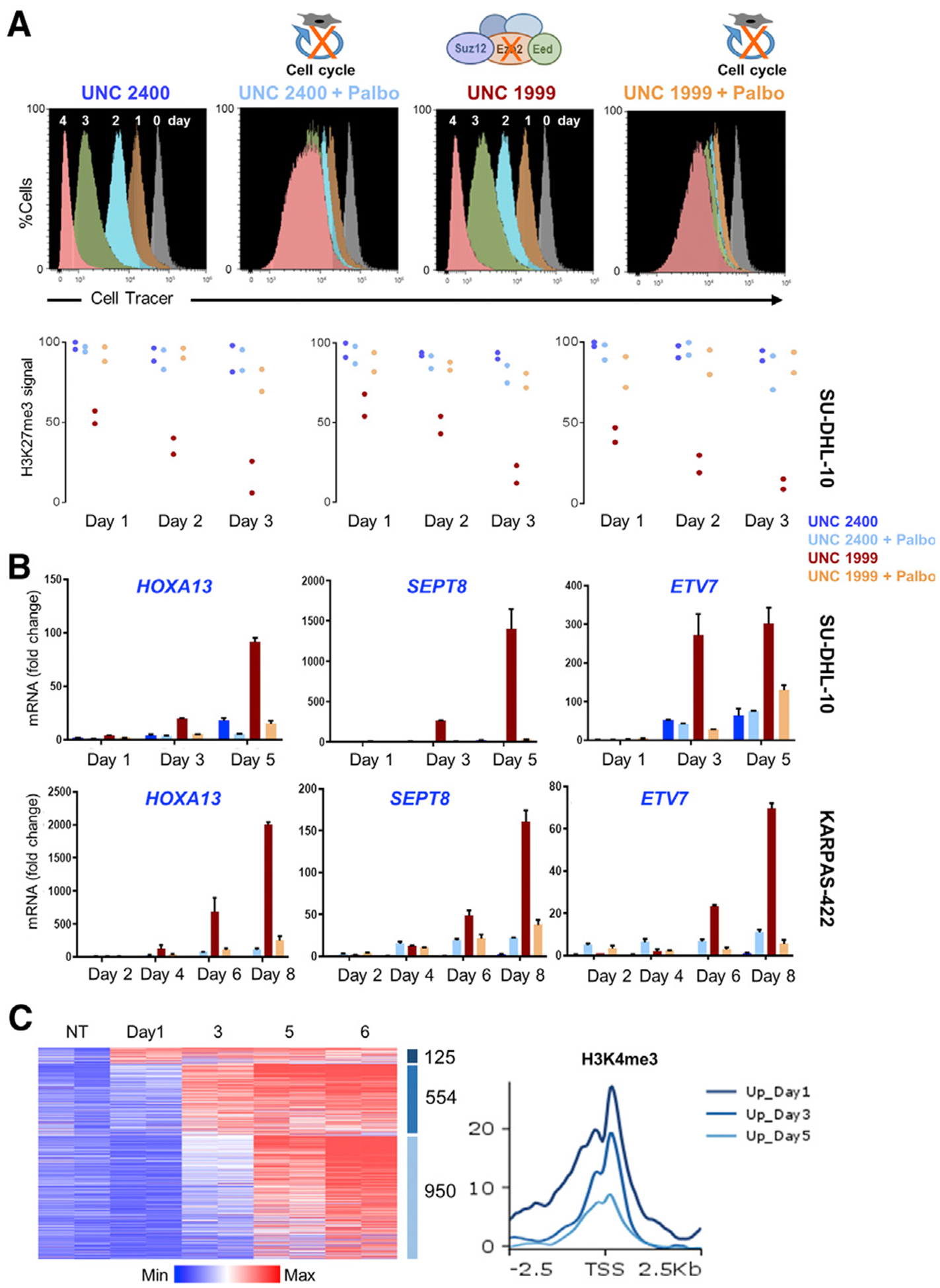
Figure 6. Cell-Replication-Dependent H3K27me3 Dilution Underlies PRC2 Target Gene Derepression in Lymphoma Cells.

(A) Histograms from flow cytometry of CellTrace-labeled SU-DHL-10 cells, showing ~50% reduced fluorescence each day, reflecting replicational dilution of the cytoplasmic dye. Cell cycle inhibitor palbociclib (Palbo) arrests this dilution, and inhibition of PRC2 by the small molecule UNC1999 or an inactive analog (UNC2400) does not materially deter cell division. qRT-PCR analysis (representing 2 or 3 biological replicates) shows gradually increasing expression of representative PRC2 target genes in UNC1999-treated SU-DHL-10 cells and attenuation of this increase in cells co-treated with Palbo. ChIP-qPCR for H3K27me3 at the corresponding promoters reveals persistent signals in control cells, ~50% reduction in UNC1999-treated cells after each division, and attenuated reduction of H3K27me3 in cells co-treated with Palbo.
(B) Likewise, PRC2 target genes increase expression only after repeated cell division in UNC1999-treated KARPAS-422 cells and Palbo attenuates this increase. Data are from 2 or 3 biological replicates.
(C) Progressive activation of PRC2 target genes, measured by RNA-seq analysis of DMSO-treated cells (controls) and on different days after UNC1999 exposure of SU-DHL-10 cells. Data (log2 RPKM+1) are shown for duplicate samples from each condition. Aggregate quantitation of H3K4me3 around promoters (TSS ± 2.5 kb) of genes expressed 1, 3, or 5 days (and after). Average H3K4me3 signals are higher at promoters that activate early.
See also Figure S4.
