
Fig. 1.
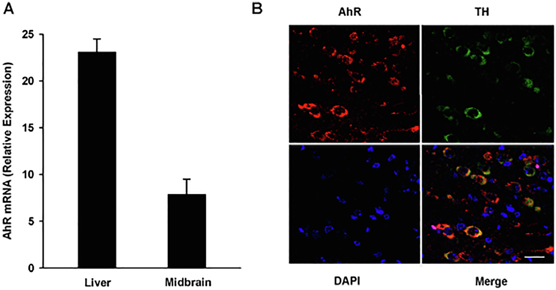
AhR is expressed in the mouse ventral midbrain and DA neurons. A) Total RNA was extracted from the ventral midbrain region and liver of sacrificed mice. AhR mRNA levels were determined by qPCR and normalized to 18S ribosomal RNA. The results are expressed as the mean ± S.D. of samples from three different mice. B) Representative confocal microscopy images of AhR expression in the ventral midbrain region. AhR, TH and DAPI were visualized as red, green, and blue, respectively. Scale bar: 25 μM.