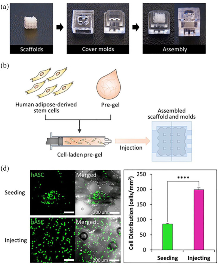
Figure 4.
(a) Assembly of cover moulds and scaffolds for cell-laden hydrogel injection. (b) Scheme of the cell-laden hydrogel pre-gel injection procedure. (c) Calcein AM staining of each scaffold after cell seeding and injecting (scale bar = 200 μm) and quantification of cell distribution in each scaffold (n = 3 per experimental group, ****p < 0.0001). (©Yi et al.67 Article distributed under a Creative Common Attribution License CC BY 4.0).

