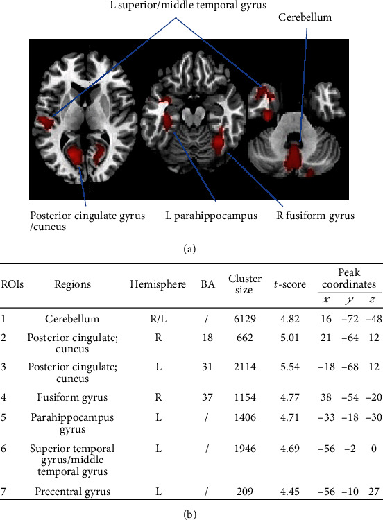
Figure 1.

Anatomical locations with significant differences in gray matter density. (a) Significant (cluster level statistics, P value < 0.05, FWE-corrected) regional gray matter volume reduction in a patient with Parkinson's disease was revealed by whole-brain VBM analysis. (b) The coordinates x, y, and z refer to the anatomical location, indicating the standard stereotactic space as defined by Montreal Neurological Institute. The t-score of the voxel with the strongest group effect in a given cluster is also listed. Abbreviations: ROIs: regions of interest; BA: Brodmann area; L: left; R: right; FWE: family-wise error; MNI: Montreal Neurological Institute; VBM: voxel-based morphometry.
