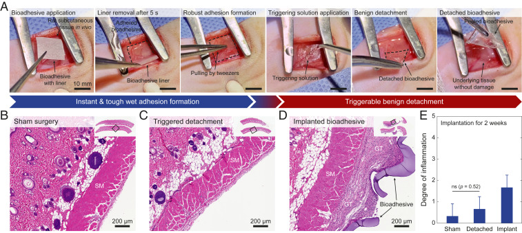
Fig. 3.
In vivo applicability and biocompatibility of the bioadhesive. (A) Photographs for instant robust adhesion and triggerable benign detachment of the bioadhesive in rat subcutaneous space in vivo. (B–D) Representative histological images stained with H&E for biocompatibility assessment of the sham surgery (B), the triggered detachment of the bioadhesive (C), and the implanted bioadhesive (D). (E) Degree of inflammation of the sham surgery, the triggered detachment of the bioadhesive, and the implanted bioadhesive groups evaluated by a blinded pathologist (0, normal; 1, very mild; 2, mild; 3, moderate; 4, severe; 5, very severe) after 2 wk of subcutaneous implantation. SM and GT indicate skeletal muscle and granulation tissue, respectively. All experiments are repeated four times with similar results. Values in E represent the mean and the SD (n = 4). P values are determined by a Student’s t test. Scale bars are shown in the images. ns, not significant (P > 0.05).

