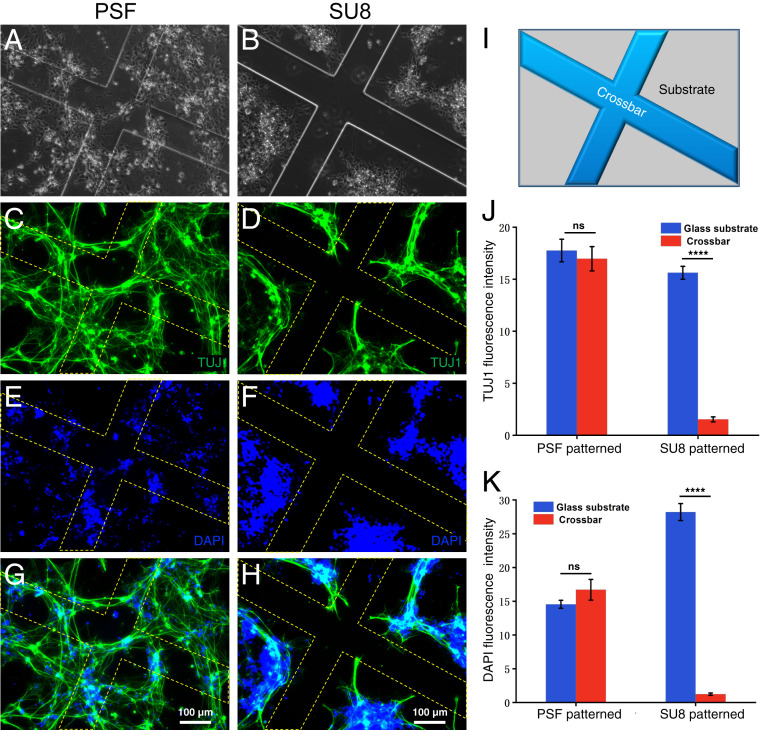
Fig. 4.
hiNSC adhesion study on microfabricated PSF and SU8 structures. (A and B) Bright-field microscope images of cells plated on (Left) PSF- and (Right) SU8-patterned surfaces. The cells were stained with neuron-specific marker antibody TUJ1 (C and D) and DAPI (E and F), with G and H merged images for both surfaces. (I) Schematic of the patterned structure. (J and K) Statistical analysis of fluorescence intensity for TUJ1 and DAPI on PSF- and SU8-patterned surfaces, showing that cells distribute evenly on the PSF-patterned surfaces, while avoiding SU8 framework (****P < 0.0001; n = 7).

