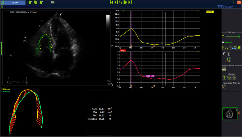
Fig. 2.
RVFAC obtained by the TomTec software (2D cardiac performance analysis) 2D speckle-tracking echocardiography. FAC was measured using RV end-diastolic area (RVDA) and end-systolic area (RVSA) obtained by manual tracing of the RV endocardium at end-diastole and end-systole in the apical 4-chamber view. The formula: [RVDA-RVSA/RVDA] × 100 was then used to calculate FAC. To limit sub-optimal interobserver reproducibility, it was ensured that the RV was contained in the whole imaging frame throughout systole and diastole, while ensuring that trabeculae in RV cavity was included. LVEF: Left ventricular ejection fraction. RVFAC: Right ventricular fractional area change. RVFWS: Right ventricular free wall strain. RVGLS: Right ventricular global longitudinal strain

