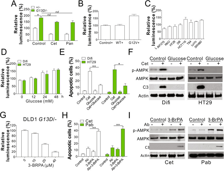
Fig. 2.
Kras mutation suppressed the AMPK activation by glycolysis. a The cellular ATP level of DLD1 WT and Kras mutation cells was analyzed by luminescence assay. b The cellular ATP level in DLD1 WT (+/−) cells stably expressing control, Kras WT, or Kras mutant (G12V) by retrovirus transfection (c) The cellular ATP level in the indicated cell lines. d The cellular ATP level of HT29 and Difi cells cultured in media containing 20 mM glucose at indicated time. e HT29 and Difi cells cultured in the media with or without 20 mM glucose were treated with Cet (15 nM for HT29; 5 nM for Difi) for 48 h. The apoptosis was analyzed by Hoechst 33258 staining. f Western blot of indicated proteins in HT29 and Difi cells treated as in (e). g The cellular ATP level of DLD1 Kras mutated treated with 10 μM 3-BrPA for indicated time points. h The apoptosis of DLD1 Kras mutated cells treated with 10 μM 3-BrPA in combined with 5 nM Cet or 10 nM panitumumab (Pab). i Western blot of indicated proteins in DLD1 Kras mutated cells treated as in (h). Each experiment was repeated for 3 times. nd, p > 0.05; *, p < 0.05; **, p < 0.01

