
Fig. 3.
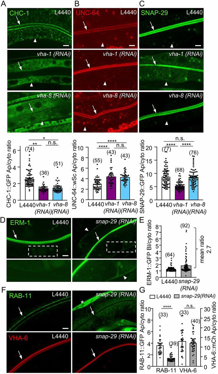
SNAP-29 acts downstream of V0-ATPase in polarity maintenance. (A-C) V0-ATPase silencing specifically decreased the apical localization of GFP::SNAP-29. Strains expressing the indicated markers were silenced for V0/vha-1 or V1/vha-8 ATPase for 72 h and then imaged. Histograms represent the quantification of the apical/cytoplasmic ratio of each marker (n=3). (D-E) snap-29(RNAi) induced a basolateral mislocalization of ERM-1. snap-29 was knocked down by RNAi in ERM-1::GFP-expressing strains and its localization was recorded after 72 h. (E) The quantification of the basolateral/cytoplasmic ratio of ERM-1::GFP (n=5). (F-G) snap-29(RNAi) also induced a RAB-11+ endosomes loss but did not affect VHA-6 apical localization. snap-29 was depleted by RNAi in a strain co-expressing GFP::RAB-11e and VHA-6::mCh. The asterisk indicates a muscular Myo-3P::GFP co-expressed with VHA-6::mCh. (G) The quantification of GFP::RAB-11e (left panel) and VHA-6::mCh (right panel) apical/cytoplasmic ratio (n=2). In all micrographs, worms are at the L4/young adult developmental stage. Arrows and arrowheads indicate the apical PM and the basolateral PM, respectively. Histograms are mean±s.e.m.; on each panel, dots represent individual worms and the total number of worms is indicated in brackets. n.s., nonsignificant, *P<0.05, **P<0.01, ****P<0.0001. Scale bars: 5 µm.