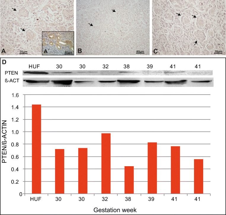
Fig. 1.
Expression of PTEN in human foetal ovary. (a–c) General view of ovarian section at 13, 18 and 30 wpc, respectively, negative for PTEN immunolocalisation both in oogonia and primordial follicles; arrows point to primordial follicles. An example of negative control staining performed by avoiding primary antibody is shown in Fig. 1S. (a’) Internal positive control showing FOXO3 expression in blood vessels from the same histological section. (d) Western blots and quantification showing low PTEN detection in all samples from 30 wpc to birth. HUF, Human uterine fibroblast cell line used as positive control (see “Materials and methods”); wpc, weeks post-conception

