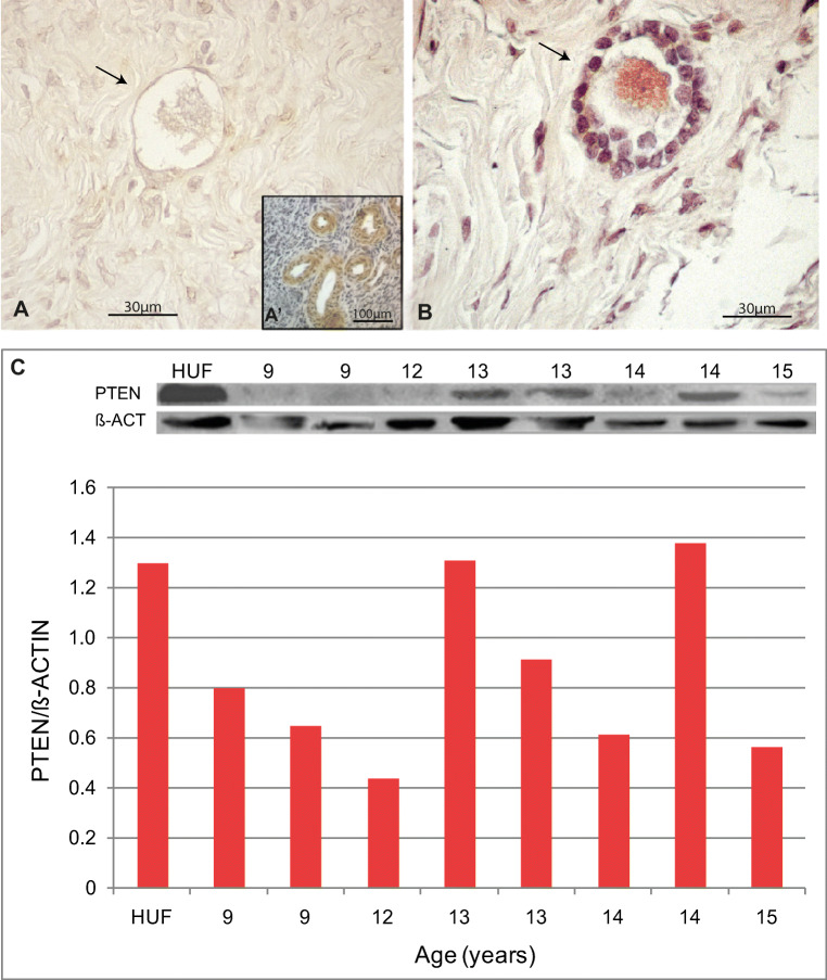
Fig. 3.
Expression of PTEN in pubertal human ovary. (a) Illustrative primordial follicle with no signal for PTEN from a 14-year-old sample. An example of negative control staining performed by avoiding primary antibody is shown in Fig. 1S. (a’) Internal positive control. (b) Unique primary-to-secondary transition follicle showing cytoplasmic localisation of PTEN protein found in the 16-year-old sample. (c) Western blot quantification showing low detection in almost all samples analysed

