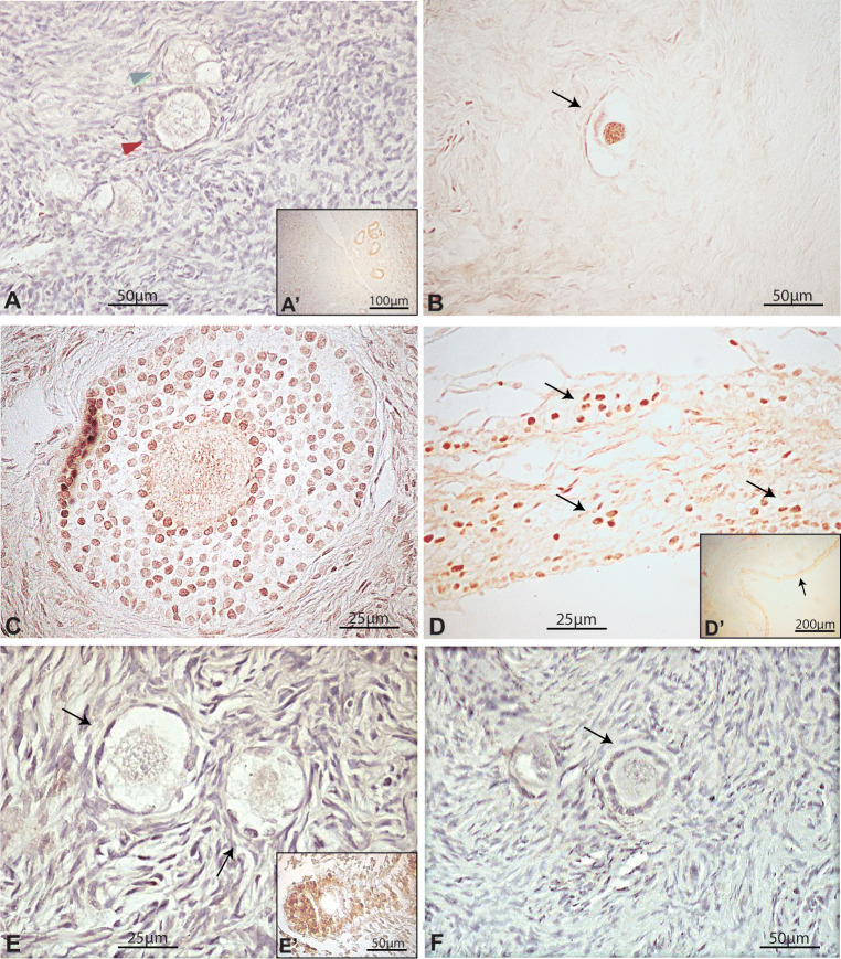
Fig. 4.
Immunohistochemical detection of FOXO3 and pFOXO3 in pubertal human ovary. (a) Pubertal ovary showing negative FOXO3 primordial (green arrowhead) and primary (red arrowhead) follicle. (b) Illustrative primordial follicle (arrow) showing nuclear FOXO3 expression. (c) FOXO3-expressing granulosa cells in pre-antral follicle. (d) Detail of FOXO-expressing granulosa cells in antral follicle; (d’) inset, arrow indicates the approximate region magnified in D. (e) Primordial follicles negative for pFOXO immunolocalosation. (f) Illustrative primary follicle negative for pFOXO immunolocalisation. Insets a´ and c´, internal positive control

