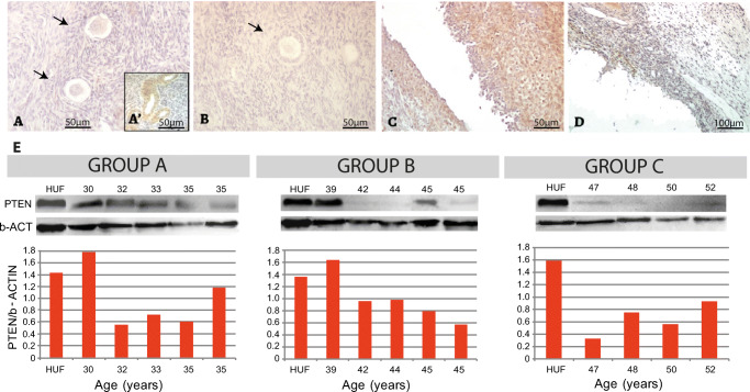
Fig. 5.
PTEN expression in the adult human ovary. (a) Illustrative example of negative PTEN expression in primordial follicles. (a’) Internal positive control, blood vessels. (b) PTEN negative staining in primary follicle. (c) Granulosa cells from an atretic follicle showing positive PTEN expression. (d) PTEN negative albicans body. (e) Western blot analysis showing low PTEN detection in almost all cases. HUF, Human uterine fibroblast cell line used as positive control (see “Materials and methods”)

