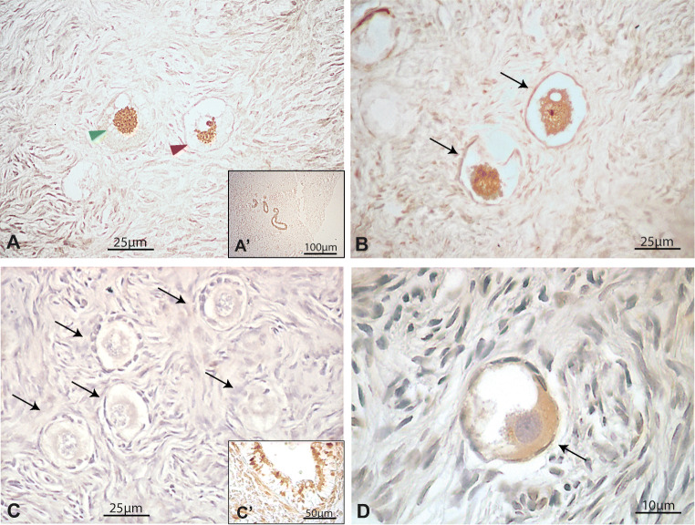
Fig. 6.
Immunohistochemical detection of FOXO3 and pFOXO3 in the human adult ovary. (a) Illustrative primordial follicles showing nuclear (green arrowhead) and cytoplasmic (red arrowhead) expression of FOXO3 and pFOXO3. (b) Nuclear FOXO3 expression in primordial follicles from a 39-year-old patient. (c) Negative primordial follicles for pFOXO3 staining. (d) Illustrative primordial follicle showing cytoplasmic localisation of pFOXO3. Insets a´ and c´, internal positive controls. An example of negative control staining performed by avoiding primary antibody is shown in Fig. 1S

