Fig. 4.

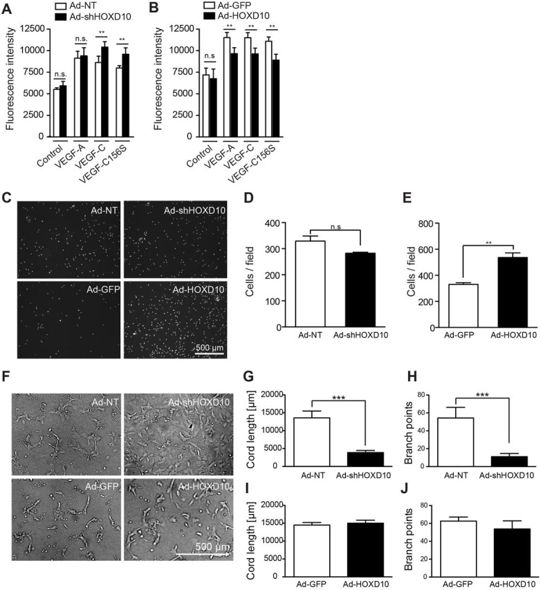
HOXD10 affects proliferation, migration and tubular morphogenesis of LECs. Proliferation of HOXD10-depleted LECs (A) and HOXD10-overexpressing LECs (B) in the absence (medium) or presence of VEGF-A, VEGF-C156S or VEGF-C. A significant increase in proliferation was observed in knockdown cells compared to control cells after treatment with VEGF-C or VEGF-C156S, whereas proliferation of HOXD10-overexpressing cells was slightly reduced. (C) Representative images of the modified Boyden chamber migration assay after 4 h. (D) After HOXD10 knockdown, LEC migration was slightly reduced (not significant). (E) HOXD10-overexpressing cells showed significantly increased migration compared to control cells. (F) Representative images of tubular morphogenesis by transduced LECs. Formation of cord-like structures in HOXD10-depleted cells was severely impaired. Instead, these cells continued to grow as a monolayer. Quantification of total cord length (G) and branch points (H) showing a strong reduction in tube formation in Ad-shHOXD10 cells compared to control (Ad-NT) cells. Cord length (I) and branch points (J) were not significantly affected by HOXD10 overexpression compared to control (Ad-GFP). Bars represent mean±s.d. (n=5 in A,B; n=3 in D,E; n=4 in G–J). **P<0.01; ***P<0.001; n.s., not significant [two-way ANOVA with Bonferroni post-test (A,B) or Student's t-test (all other panels)].
