Fig. 1.
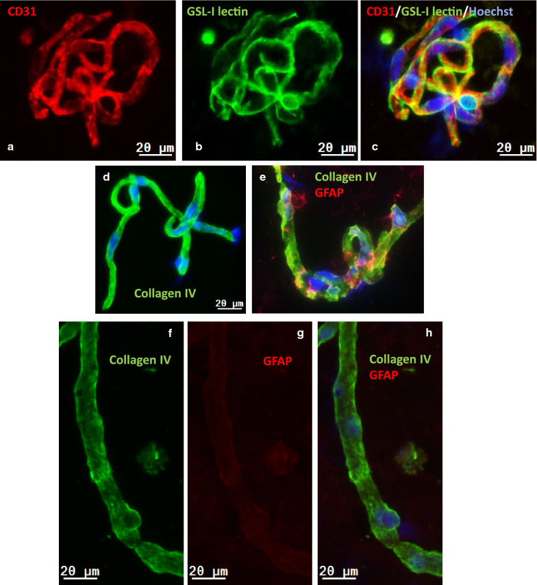
Immunofluorescence analysis of isolated brain microvessels and capillaries (BMV) from the mouse (a–e) and human (f–i) brain. a CD31 immunofluorescence (red) in mouse BMV; b GSL-I lectin staining (green) in mouse BMV. c A composite image of CD31, GSL-I and Hoechst-stained nuclei (blue) of the same BMV. d Collagen IV immunofluorescence (green) in mouse BMV. e A composite image of collagen IV (green) and GFAP (red) immunofluorescence and Hoechst-stained nuclei (blue) in mouse BMV. f Collagen IV immunofluorescence of human BMV. g GFAP immunofluorescence of human BMV. h A composite image of of collagen IV (green) and GFAP (red) immunofluorescence and f Hoechst-stained nuclei (blue) in human BMV. i Hoechst staining of nuclei in human BMV. The scale bar is 20 µm

