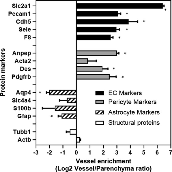
Fig. 2.

The enrichment of the blood–brain barrier (BBB)-related proteins in mouse brain microvessels (BMV) compared to brain parenchyma. BMVs and vessel-free brain parenchyma were isolated as described in Materials and Methods and relative enrichment of key protein markers in each fractions was determined by proteomics. Shown are Log2 ratios between levels of various proteins in purified vessels and vessel-depleted parenchyma. Protein markers correspond to: (1) Endothelial cell markers: Slc2a1, Glucose transporter (Glut1); F8, Coagulation factor VIII-related antigen; Sele, E-selectin; Cdh5, VE-cadherin; Pecam1, CD31; (2) Pericyte markers: Pdgfrb, Platelet-derived growth factor receptor beta; Des, Desmin; Acta2, smooth muscle actin; Anpep: CD13 pericyte marker (Aminopeptidase N); (3) Astrocyte markers: Gfap, Glial fibrillary acidic protein; S100β, Protein S100-beta; Slc4a4, Electrogenic sodium bicarbonate cotransporter 1; Gja1, Gap junction alpha-1 protein; Aqp4, Aquaporin-4; (4) Structural proteins: Actb, Cytoplasmic actin; Tubb1, beta tubulin. Error bars represent mean ± SD of three biological replicates
