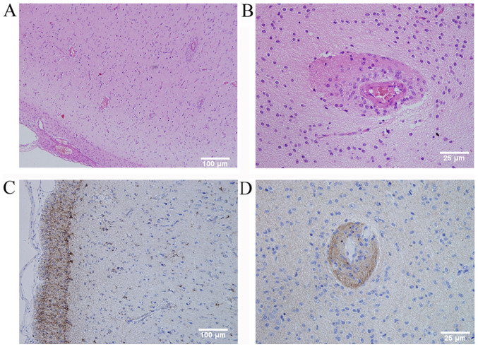
Figure 2.
(Case 1) (A and B) Histopathological examination revealed that tumor cells surrounded the blood vessels and neurons in the cortex (H&E staining; original magnification, ×100 in A and ×400 in B; scale bar, 100 µm in A and 25 µm in B). (C) Immunohistochemical staining demonstrated cytoplasmic immunoreactivity for GFAP (original magnification, ×100; scale bar, 100 µm) and (D) dot-like staining for EMA (original magnification, ×400; scale bar, 25 µm). GFAP, glial fibrillary acidic protein; EMA, epithelial membrane antigen; H&E, hematoxylin and eosin.

