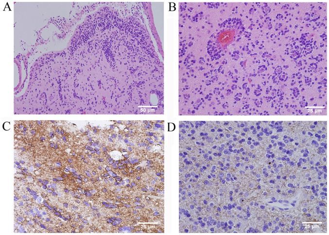
Figure 5.
(Case 2) Histopathological examination revealed infiltrative round or ovoid tumor cells in and under the cortex, and partly arranged around blood vessels and neurons in concentric sleeves and pseudorosettes and demonstrated an angiocentric and creeping pattern (H&E staining; original magnification, ×200 in A and ×400 in B; scale bar, 50 µm in A and 25 µm in B). (C) Immunohistochemical staining demonstrated strong cytoplasmic immunoreactivity for GFAP (original magnification, ×400; scale bar, 25 µm) and (D) dot-like staining for EMA (original magnification, ×400; scale bar, 25 µm). GFAP, glial fibrillary acidic protein; EMA, epithelial membrane antigen; H&E, hematoxylin and eosin.

