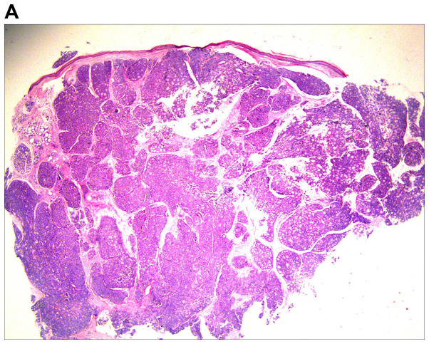
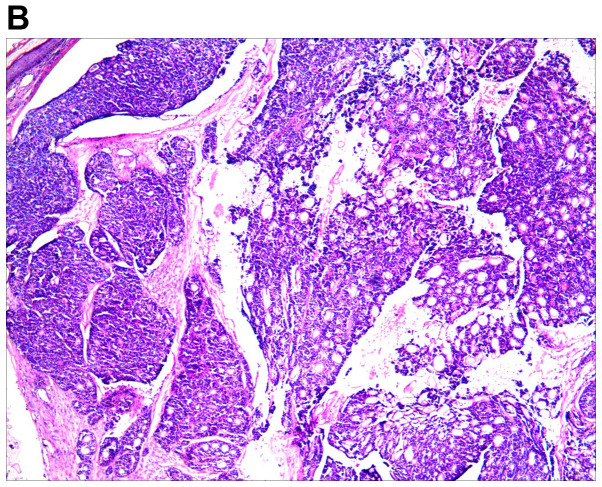
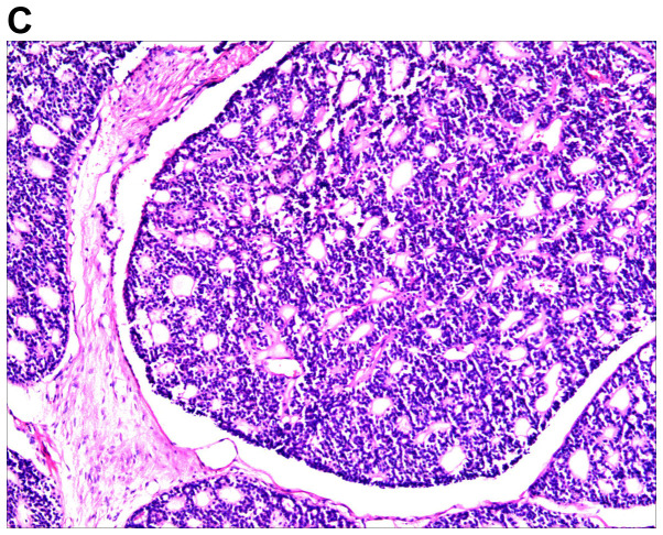
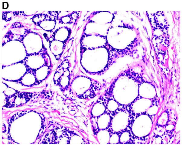
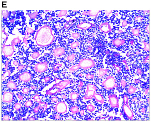
Figure 2.
Histological images of the tumor. (A) Histopathology revealed a cellular tumor with sharp circumscription, and ductal differentiation and focal myxoid stroma at low power (H&E; magnification, ×12.5). (B-D) The tumor cells exhibited a tubular and characteristic cribriform growth pattern, with hyaline material within and around the tumor cells. The tumor cells appeared uniform with vesicular nuclei and prominent nucleoli. Clear cell forms and cystic spaces are evident (H&E; magnification, ×25 in B, ×100 in C, and ×200 in D). Histological images of the tumor. (E) The cystic spaces are highlighted by the periodic acid-Schiff stain (magnification, ×200).

