Figure 4.
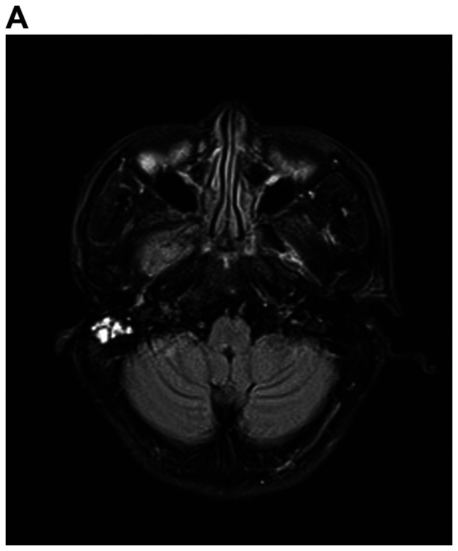
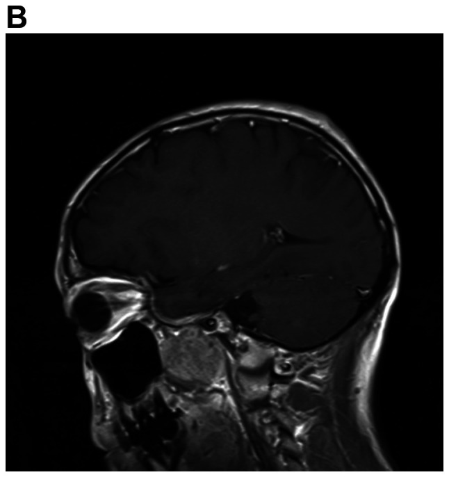
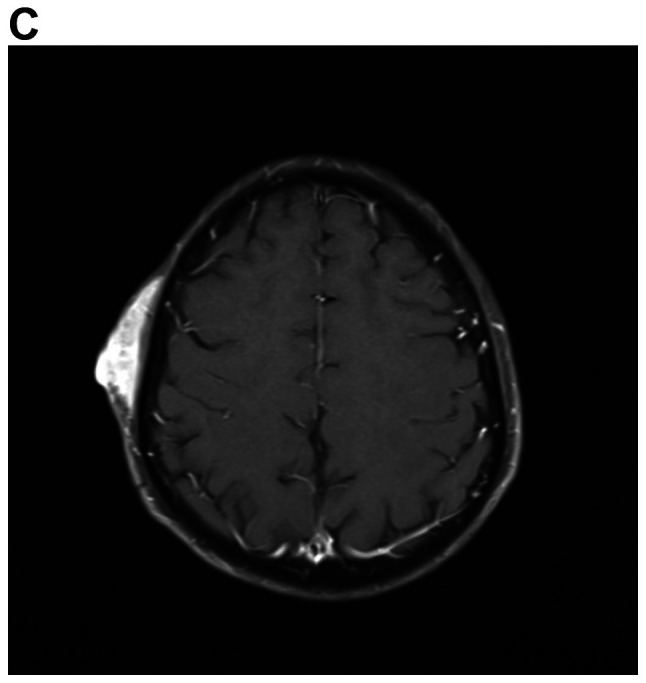
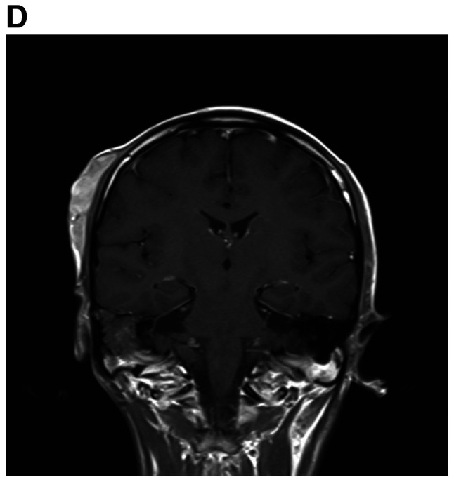
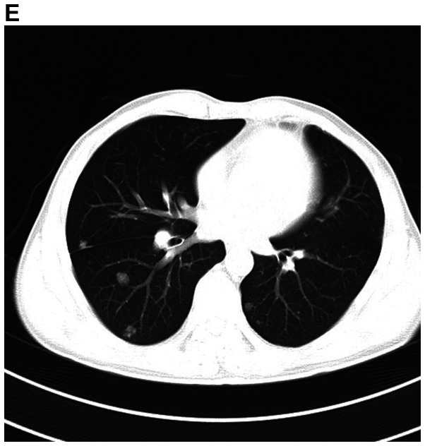
MRI demonstrates superior contrast resolution of multiple mass lesions in the right pharynx, inferior temporal fossa, middle cranial fossa and right frontotemporal region. (A and B) T2WI. (A) Coronal scan and (B) sagittal scan. In the right mastoid process, an enhanced T2 signal suggested inflammation of the right mastoid process. (C and D) On T1WI, the right side of the temporal region featured an enhanced T1 signal suggesting the presence of a subcutaneous neoplasm. (C) Coronal scan and (D) sagittal scan. MRI demonstrates superior contrast resolution of multiple mass lesions in the right pharynx, inferior temporal fossa, middle cranial fossa and right frontotemporal region. (E) Chest enhanced CT scan indicated multiple metastases in the bilateral lungs. T1WI, T1-weighted imaging.

