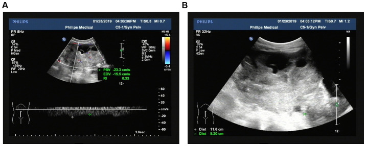
Figure 1.
Ultrasonic acoustic and blood flow display images of ovarian cancer. (A) Ultrasonic acoustic image of ovarian cancer. (B) Blood flow display image of ovarian cancer. Ovarian cancer: the size of the tumor was 11.6×9.2×10.2 cm, the shape was irregular, the boundary was unclear, the boundary was irregular and solid was the main feature. CDFI of its internal detection and rich blood flow signal and RI was 0.33. CDFI, color doppler flow imaging; RI, resistance index.

