Figure 3.

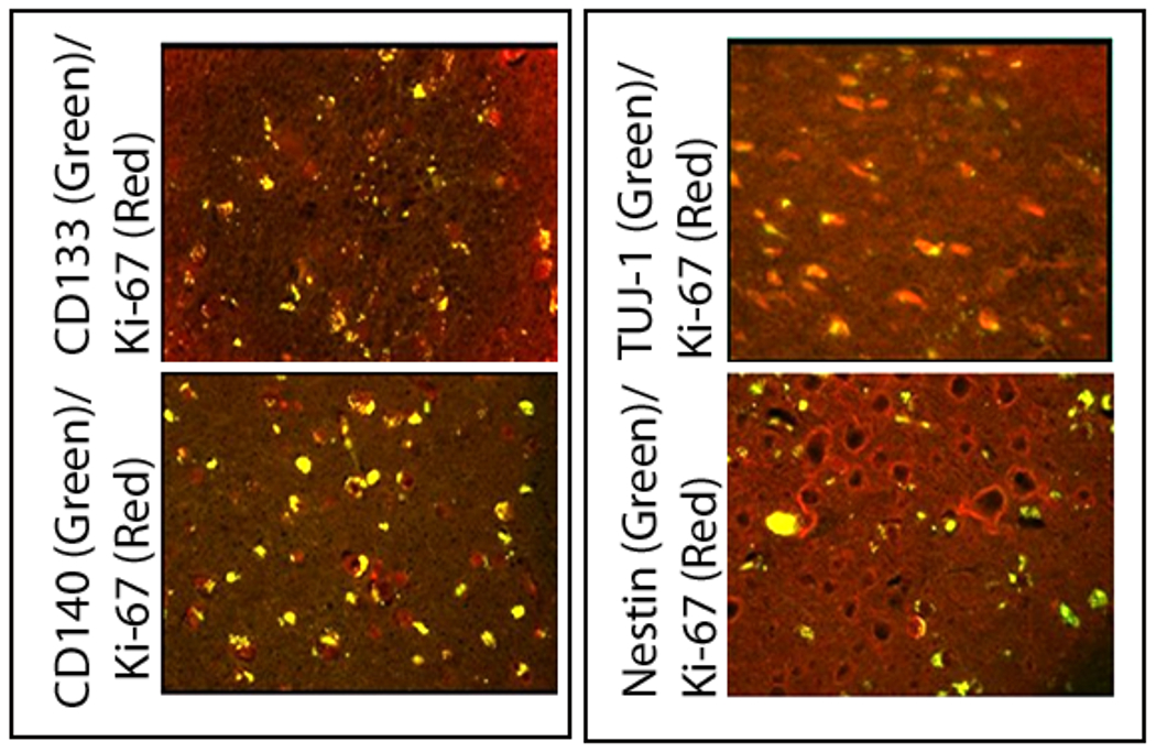
Confocal photomicrographs of left frontal GB recurrence (Case #1) showing the co-labeling of cells (yellow) with Ki-67 and CD133 (top left), CD140 (bottom left), TUJ-1 (top right) and nestin (bottom right). This demonstrates that cells labeled with several different types of progenitor markers found in and around the GB recurrence are actively dividing.
