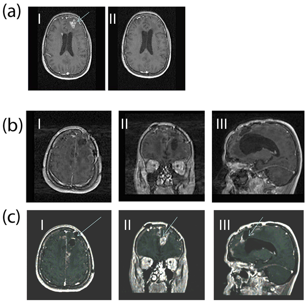
Figure 4.

(a) Gadolinium enhanced MR images (Case 1) comparing pre- (I) and post-DepoCyt treatment (II) after completion of the consolidation phase (cycle 6). After four months of treatment, the original left frontal lesion, and periventricular enhancement has disappeared. (b) MRI 40 weeks after restarting ITV DepoCyt in patient (Case #1). Images (I, II, III) demonstrate a marked decrease in the bihemispheric tumor burden as compared to Figure 4c. (c) MRI 6 weeks after discontinuing ITV DepoCyt in patient (Case#1). Bihemispheric recurrent GB is demonstrated (I, II, III).
