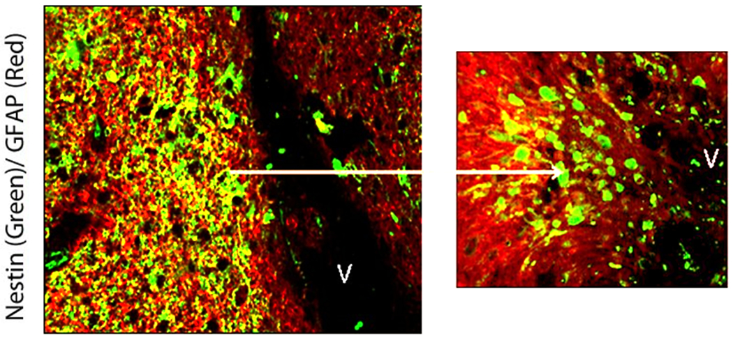
Figure 6.

Confocal photomicrographs of right frontal periventricular GB recurrence in patient #2. Nestin immunoreactivity (green) and GFAP immunoreactivity (red) are demonstrated in and around the GB recurrence. Nestin positive cells are more abundant closer to the SVZ nearest to the dorsolateral horn of the right lateral ventricle (V). Nestin+ cells are seen streaming from the ventricular surface towards the GB recurrence (left picture). Co-labeling of GFAP+/Nestin+ (yellow) cells appear in these regions.
