
Figure 1. Oc, Ets65A and Sox102F are expressed in T1 neurons.
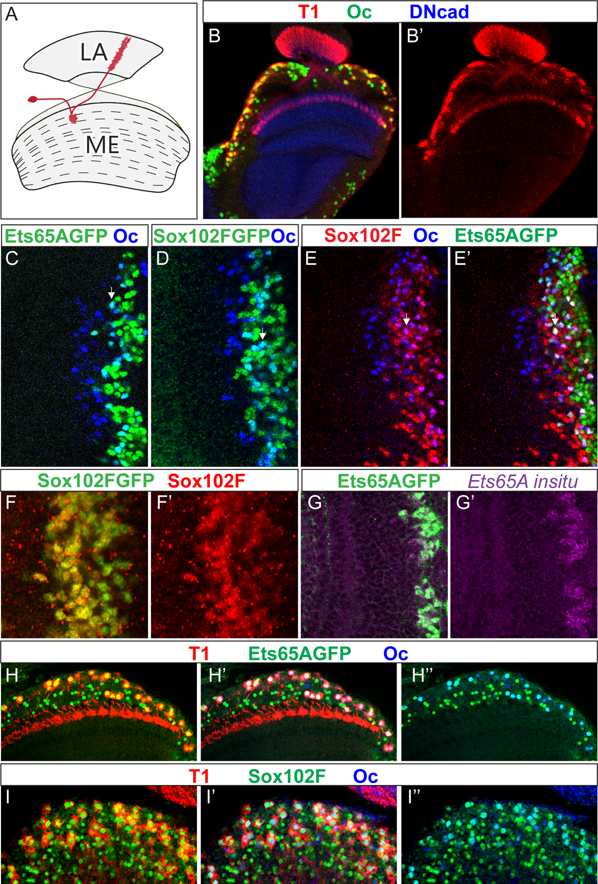
(A) A schematic drawing of the normal T1 morphology in red. LA: lamina. ME: Medulla. (B-B’) The expression of Oc (green) in the adult medulla carrying T1LexA>LexopRFP (red). (C,D) The expression of Ets65A::GFP (green in C) or Sox102F::GFP (green in D) partially overlaps with that of Oc (blue) in the 3rd instar larval medulla. The early-born (deeper layer) neurons are to the left, and later born (more superficial layers) neurons are to the right. Neurons expressing both appear cyan in the overlay. White arrow indicates one example. (E,E’) Neurons that express both Sox102F (red) and Oc (blue) also express Ets65A::GFP (green). (F,F’) Sox102F::GFP (green) is expressed in the same neurons as Sox102F immunostaining (red) in the 3rd instar larval medulla. (G,G’) In the 3rd instar larval medulla, the expression of Ets65A::GFP (green) is in the nucleus of the same cells that express Ets65A mRNA as shown by in situ hybridization against all isoforms (purple). (H–H”) T1 neurons (red) in the adult medulla express both Ets65A::GFP (green) and Oc (blue). (I,I’) T1 neurons (red) in the adult medulla express both Sox102F (green) and Oc (blue).